February 21st 2020 11:30, room U2-03
Dr. Alberto Bravin
European Synchrotron Radiation Facility, ID17 Biomedical beamline, Grenoble, France
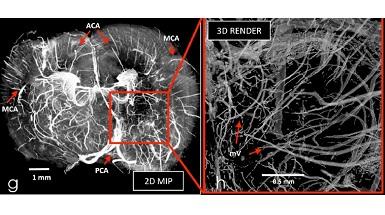
Synchrotron radiation facilities are laboratories where the most intense and collimated X-ray beams on Earth are made available to researchers for a wide range of applications. In medicine, synchrotron Xrays are used to develop new imaging, radiation therapy and surgery techniques, applied to in-vitro and in-vivo models. The high degree of coherence of the X-ray issued by a synchrotron source permit to apply powerful imaging techniques like phase contrast computed tomography imaging (PCI-CT). This is an experimental methodology, which simultaneously provides micrometric spatial resolution, high soft-tissue sensitivity and full-organ coverage without any need for sample dissection, staining/labelling or contrast agent injection.
A platform for stereotactic radiation therapy trials targeting brain tumours and a station for preclinical investigations using high dose rate (FLASH) beam therapy using microbeams are made available to the international users’ community. In the first case, beams can be stereotactically delivered to a tumour previously loaded with a high-Z material that allows to locally enhancing the dose deposition. In microbeam radiation therapy, multienergy X-rays (dose rate >10,000 Gray/s) are spatially fractionated in arrays of microscopic beams (25-600 μm wide, 50 to 1200 μm spaced) and delivered with submillimetric precision. Doses up to hundreds of Grays, delivered in a fraction of a second, can be very well tolerated by the tissues as shown in several small and large animal models.
After a brief introduction to synchrotron radiation, highlights on the application of PCI-CT in bioimaging and the state-of-the-art in radiotherapy and radiosurgery using synchrotron radiation will be presented.