Nanoscopy for biotechnology and Medicine

Nanoscopy is a branch of optical microscopy that overcomes the optical limitations set by diffraction of radiation. This limitations, as developed by Abbe, states that the resolution obtained by means of a radiation of wavelength λ is given by the wavelength and the numerical aperture, NA, of the collecting optics:

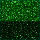
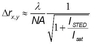
Nanoscopy tries to overcome this limitation by exploiting time and space structured illumination and spectroscopy. The group is involved in research related to the so called STED microscopy in which two superimposed laser spots (a Gaussian and a Toroidal spot) raster scan the sample. This technique exploits the stimulated emission of the fluorophores (induced by the toroidal spot induced by the so called STED laser) in the periphery of the conventional Gaussian spot that primes the fluorescence emission. The laboratory is equipped with a CW-STED Leica microscope. With the STED microscopy the space resolution in the focal plane increases as the square root of the intensity of the toroidal (STED laser) spot:

Funding
Università di Milano Bicocca, Fondo Grandi Attrezzature.
Collaborations
- University of Parma: S. Bettati, A. Mozzarelli;
- Humanitas: C. Rumio;
- I.Rivolta, Dept. Exp. Medicine; University of Milano Bicocca;
- E. Martegani, Dept. Biotechnology; University of Milano Bicocca.
Contact
Publications
G. Chirico, Occhiali Nuovi per le Nano-Biotecnologie, Il Mondo del Laboratorio, Aprile 2011: 29-31.