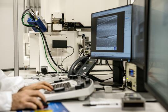
Optical microscopy and spectroscopy laboratory
Contact person: Prof. Maddalena Collini
Service info
Image

- The standard spectroscopic characterization of protein samples includes UV-VIS absorption, fluorescence emission and excitation, near-UV and far-UV circular dichroism.
- The dimension and the aggregation state of proteins and nanoparticles are assessed by means of polarized (90 deg) and/or depolarized (different angles) light scattering.
- The spectroscopic characterization of fluorescent molecules and nanoparticles with non linear excitation comprises the possibility to measure the two-photon excitation spectrum (from 700 nm to 980 nm), the emission spectrum by means of a CCD, and the diffusion and photodynamic by means of fluorescence correlation spectroscopy.
- The cellular uptake of nanoparticles and nanostructures is studied by means of non linear excitation microscopy (700-980 nm) and its characterization involves time resolved imaging experiments at different nanosystem concentrations, to assess both the uptake and the toxicity. FRAP (fluorescence recovery after photobleaching) measurements can be exploited to investigate the intracellular diffusion.
- Confocal and super-resolution imaging are based on a Leica TCS CW STED microscope: excitation wavelengths 458 nm, 476 nm, 488 nm, 514 nm, 561 nm, 633 nm. Detectors: 2 PMT + 2 Hybrid. Scan speed from 200 to 1400 Hz, 8000 Hz in resonant scanning mode. Image modes: xyz, xyt, xyzt, linescan, mosaic, spectral.
Analysis pricelist
Our analysis pricelist.
(Pricelists are in Italian only, but you can contact one of the staff member for details).
Analysis timetable
Analysis timetable will depend on the desired service and will be agreed at the moment of the request.
Results
You will receive results by e-mail together with a report about equipment, set-up, techniques and measurement accuracy.
More information
Prof. Maddalena Collini maddalena.collini@unimib.it Tel. 02 6448 2439